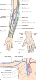
Figure 17.1
(a) Superficial veins of the forearm. (b) Superficial veins of the dorsal aspect of the hand. (c) Central veins and veins of the upper arm.

Figure 17.5
Chest X‐ray showing catheter tip correctly positioned (arrow).

Figure 17.9
Vessel Health Preservation (VHP) framework. Source : Reproduced from Hallam et al. ( ) with permission of Sage Publications.

Figure 17.13
Ported cannula.

Figure 17.17
Visual Infusion Phlebitis (VIP) scale. Source : Reproduced from Jackson ( ) with permission of Nursing Times .
Figure 17.21
Documentation example from The Royal Marsden NHS Foundation Trust's vascular access device documentation booklet. Source : Reproduced with permission...

Figure 17.25
PICC Zone Insertion Method (ZIM). Source : Reproduced from Dawson ( ) with permission of Elsevier.

Figure 17.29
Example peripherally inserted central catheter (PICC) consent form.
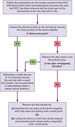
Figure 17.33
Algorithm for the removal of a peripherally inserted central catheter (PICC). Source : Adapted from Hughes ( ).

Figure 17.37
StatLock securement device.

Figure 17.41
Implanted ports.

Figure 12
Turn the tap to close off the pre‐filled syringe and open it to the empty syringe.

Figure 19
Checking the needle tip.

Figure 28
Disposing of the stylet into a sharps bin.

Figure 10
Apply gel to the area and, using the ultrasound probe, assess and select the vein.

Figure 24
Flashback into the cannula chamber when the vein is punctured.

Figure 35
Semi‐permeable transparent IV film dressing.

Figure 24
Advancing the introducer.

Figure 31
Advancing the introducer.

Figure 20
Positioning, securing and labelling the cannula.

Figure 17.2
The main veins used for central venous access device placement. Source : Reproduced from Dougherty ( ) with permission of John Wiley & Sons.

Figure 17.6
Electrocardiogram (ECG) pattern indicating raised P waves.

Figure 17.10
Patient information booklet on central venous access devices.

Figure 17.14
Safety non‐ported cannula.

Figure 17.18
Cleaning the skin.

Figure 17.22
Applying an ultrasound probe to the arm to locate veins.
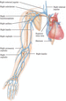
Figure 17.26
Anterior view of the superficial veins.
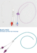
Figure 17.30
Peripherally inserted central catheter (PICC) types.

Figure 17.34
Non‐tunnelled multilumen central venous catheter.
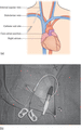
Figure 17.38
Tunnelled catheter. (a) Anatomical positioning of tunnelled catheter. (b) Patient with tunnelled catheter in situ .

Figure 17.42
Non‐coring needles have the penetration style of a knife so when the needle is removed, the septum closes behind it.

Figure 14
Unblocking an occluded catheter. (a) Aspirate on an empty syringe, which creates negative pressure. (b) Turn the tap to close off the empty syringe an...

Figure 21
Inserting the cannula and waiting for first flashback. (a) Open cannula. (b) Integrated closed system cannula.

Figure 29
Flushing the cannula. (a) Open cannula. (b) Integrated closed system cannula.

Figure 19
Using aseptic non‐touch technique, apply sterile gel to the transducer on the ultrasound probe and cover it with a sterile semi‐permeable transparent ...

Figure 25
Ultrasound image of the cannula inside the vein.

Figure 18
Wire being threaded in the cannula.

Figure 25
Wire being threaded in the cannula.
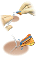
Figure 44
Attaching the securing device to the skin. Source : Reproduced with permission of Interrad Medical, Inc.

Figure 17.3
Clot formations. Source : Reproduced from Macklin and Chernecky ( ) with permission of Elsevier.

Figure 17.7
SecurAcath securing device. Source : Reproduced with permission of Interrad Medical, Inc.

Figure 17.11
(a) Vein visualization device. (b) Vascular access ultrasound scanner.

Figure 17.15
Safety integrated closed system.
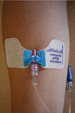
Figure 17.19
Peripheral cannula secured with StatLock.

Figure 17.23
Ultrasound images of veins on screen.
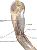
Figure 17.27
Anteromedial view of the superficial veins of the arm and forearm. Source : Reproduced from Tortora and Derrickson ( ) with permission of John Wiley ...

Figure 17.31
Peripherally inserted central catheter (PICC) with adhesive securing device.

Figure 17.35
Types of catheter tip. (a) Open‐ended catheter (single and double lumen). (b) Staggered‐exit open‐ended catheter.

Figure 17.39
Groshong two‐way valve catheter. (a) Infusion (positive pressure). (b) Aspiration (negative pressure). (c) Closed (neutral pressure).

Figure 17.43
Normal arterial trace.

Figure 10
Palpating the vein.

Figure 24
Second flashback. (a) Open cannula. (b) Integrated closed system cannula.

Figure 30
Method of taping a peripheral cannula.

Figure 21
Apply sterile gel and using the non‐dominant hand position the ultrasound probe 0.5–1.0 cm above the proposed site of insertion.

Figure 26
Withdraw the stylet while advancing the rest of the cannula.

Figure 21
Local anaesthetic injection.

Figure 28
Local anaesthetic injection.

Figure 8
Removing the SecurAcath device. Source : Reproduced with permission of Interrad Medical, Inc.

Figure 17.4
(a) Algorithm for partial withdrawal occlusion – that is, fluids can be infused freely by gravity but blood cannot be withdrawn from the catheter. (b)...

Figure 17.8
Venous Assessment Tool (VAT) score. Source : Reproduced from Wells ( ) with permission of Nursing Standard .
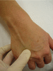
Figure 17.12
Anchoring the vein with the thumb. Source : Reproduced from Dougherty ( ) with permission of John Wiley & Sons.

Figure 17.16
I‐DECIDED: IV Assessment and Decision Tool. Source : Reproduced from Ray‐Barruel et al. ( ) with permission of BMJ .

Figure 17.20
(a) Cannula in situ . (b) Cannula secured with a semi‐permeable transparent IV film dressing.

Figure 17.24
Ultrasound cross‐sectional image of the right internal jugular vein (IJV) without compression through the probe. Image orientation as seen from the he...

Figure 17.28
Electrocardiogram (ECG) tracing showing P wave changes depending on catheter tip position.

Figure 17.32
Royal Marsden NHS Foundation Trust algorithm for the management and treatment of CVAD‐related thrombosis. CVAD, central venous access device; LMWH, lo...

Figure 17.36
One way to achieve the Trendelenburg position.

Figure 17.40
Implantable port cross‐section, accessed with non‐coring needle.

Figure 17.44
The Allen test.

Figure 14
Opening the equipment.

Figure 27
Applying digital pressure and removing the stylet.

Figure 33
Semi‐permeable transparent IV film dressing.

Figure 22
Puncture through the skin, 0.5–1.0 cm below the probe, at the selected angle.

Figure 32
Method for taping a peripheral cannula.

Figure 22
Making an incision with a scalpel.

Figure 29
Making an incision with a scalpel.

Figure 10
Flushing a port.