You are viewing a javascript disabled version of the site. Please enable JavaScript for this site to function properly.
Go to headerGo to navigationGo to searchGo to contentsGo to footer
Go to chapter navigation

Figure 14.26
Blood pressure management

Figure 14.4
Pulse points. Source : Reproduced from Tortora and Derrickson ( ) with permission of John Wiley & Sons.

Figure 14.8
Posterior tibial pulse. Source : Reproduced from Peate and Wild ( ) with permission of John Wiley & Sons.

Figure 14.13
A normal electrocardiogram (ECG) (lead II). P wave, arterial depolarization; QRS complex, onset of ventricular depolarization; T wave, ventricular rep...

Figure 14.17
Electrocardiogram (ECG) calibration verification signal. Source : Reproduced from Crawford and Doherty ( ) with permission of MA Healthcare.

Figure 14.21
Blood pressures in various parts of the cardiovascular system. The white line is the mean (average) blood pressure in the aorta, arteries and arteriol...
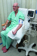
Figure 14.25
Correct blood pressure reading technique.

Figure 14.30
Factors that influence rate and depth of breathing. Source : Marieb and Hoehn ( ).

Figure 14.35
Normal peak expiratory flow rate measurements. Source : Reproduced with permission of Clement Clarke International, Ltd ( www.peakflow.com ).

Figure 14.40
Reasons for predisposition to urinary tract infections (UTIs).

Figure 14.47
The cerebrum. Source : Reproduced from Tortora and Derrickson ( ) with permission of John Wiley & Sons.
Figure 14.51
Glasgow Coma Scale and neurological observations chart.

Figure 14.36
Manual peak flow meter technique.
Figure 14.1
National Early Warning Scoring System 2 (NEWS 2). Source : RCP ( ).

Figure 14.5
Factors that increase cardiac output. Source : Reproduced from Tortora and Derrickson ( ) with permission of John Wiley & Sons.

Figure 14.9
Popliteal pulse. Source : Reproduced from Peate and Wild ( ) with permission of John Wiley & Sons.

Figure 14.14
Einthoven triangle.

Figure 14.18
Right‐sided chest lead positioning for an electrocardiogram (ECG).

Figure 14.22
The renin–angiotensin–aldosterone system. Source : Reproduced with permission of Soupvector – Own work, CC BY‐SA 4.0 https://commons.wikimedia.org/w...

Figure 14.27
Pressure changes in pulmonary ventilation. Source : Reproduced from Tortora and Derrickson ( ) with permission of John Wiley & Sons.

Figure 14.31
Spirogram of lung volumes and capacities. The average values for a healthy average male and female are indicated, with the values for a female in pare...

Figure 14.37
Mechanisms of body temperature regulation.

Figure 14.41
Significant bacteriuria. Specimens of urine are rarely sterile. A cut‐off point is identified to distinguish true infection (significant bacteriuria) ...

Figure 14.48
External anatomy of the spinal cord and the spinal nerves (posterior view). Source : Reproduced from Tortora and Derrickson ( ) with permission of Jo...

Figure 14.52
Normal and abnormal flexion and extension.

Figure 14.43
Take a blood sample from the side of the finger using a lancet, ensuring that the site of piercing is rotated.

Figure 14.2
Situation‐Background‐Assessment‐Recommendation (SBAR) tool.

Figure 14.6
Conduction system of the heart. Auto‐rhythmic fibres in the SA node, located in the right atrial wall, act as the heart's pacemaker, initiating cardia...

Figure 14.11
A 12‐lead electrocardiogram (ECG). Source : Reproduced from https://meds.queensu.ca/central/assets/modules/ECG/the_12_lead_ecg.html .

Figure 14.15
Position of chest electrodes for a 12‐lead electrocardiogram (ECG). Source : SCST ( ) – The Society for Cardiological Science and Technology ( www.sc...

Figure 14.19
(a) Normal electrocardiogram tracing. Abnormal electrocardiogram tracings: (b) first‐degree AV block, (c) atrial fibrillation, (d) ventricular tachyca...

Figure 14.23
Arterial line and transducer set.

Figure 14.28
Summary of events of inhalation and exhalation. Source : Reproduced from Peate and Wild ( ) with permission of John Wiley & Sons.
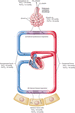
Figure 14.32
Changes to partial pressures of oxygen and carbon dioxide (in mmHg) during internal and external respiration. Source : Reproduced from Tortora and De...

Figure 14.38
Tympanic membrane thermometer.
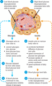
Figure 14.42
Negative feedback regulation of the secretion of glucagon and insulin Source : Reproduced from Tortora and Derrickson ( ) with permission of John Wil...

Figure 14.49
Trapezium squeeze.
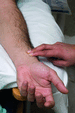
Figure 14.10
Taking a radial pulse

Figure 14.44
Insert the test strip into the blood glucose monitor and apply the blood to the test strip. Ensure that the window on the test strip is entirely cover...

Figure 14.3
Taking a radial pulse. Source : Reproduced from Peate and Wild ( ) with permission of John Wiley & Sons.

Figure 14.7
Dorsalis pedis pulse. Source : Reproduced from Peate and Wild ( ) with permission of John Wiley & Sons.

Figure 14.12
Three‐lead electrocardiogram (ECG) electrode placement.

Figure 14.16
Mason–Likar 12‐lead electrocardiogram (ECG) system.

Figure 14.20
Factors that lead to an increase in blood pressure. Changes noted within green boxes increase cardiac output, whereas changes noted within blue boxes ...

Figure 14.24
Korotkoff sounds.
Figure 14.29
Negative feedback mechanisms by which changes in PaCO 2 and blood pH regulate ventilation. Source : Marieb and Hoehn ( ).
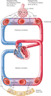
Figure 14.33
Transport of oxygen (O 2 ) and carbon dioxide (CO 2 ) in the blood. Source : Reproduced from Tortora and Derrickson ( ) with permission of John Wiley...

Figure 14.39
Summary of filtration, reabsorption and secretion in the nephron and collecting duct. Source : Reproduced from Tortora and Derrickson ( ) with permis...

Figure 14.46
The brain. Source : Reproduced from Peate and Wild ( ) with permission of John Wiley & Sons.

Figure 14.50
Pupil size guide.

Figure 14.34
Position of an oxygen saturation probe.
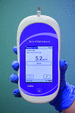
Figure 14.45
Read the result.