Figure 12.33
Chest drain: Removal

Figure 12.4
Nasal cannula.

Figure 12.8
Tracheostomy mask.

Figure 12.12
Heat and moisture exchanger for a laryngectomy.

Figure 12.16
Non‐invasive ventilation (NIV) helmet.

Figure 12.20
A chest drain and underwater seal bottle are used to drain a left pleural effusion.

Figure 12.24
Underwater chest drainage system.

Figure 12.28
Mattress suture.
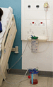
Figure 12.32
Chest drain on suction.

Figure 12.37
Tracheal dilator.

Figure 12.41
Portex® Blue Line Ultra® Sucationaid tracheostomy tube.

Figure 12.45
Portex® Uniperc® adjustable flange tracheostomy tube with Soft Seal® cuff and inner cannula.

Figure 12.49
Tracheal suction using a fine‐bore suction catheter.

Figure 12.53
Decannulation plug.

Figure 12.57
Assessing for signs of life.
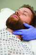
Figure 12.61
Feeling the carotid pulse.

Figure 12.65
Two people ventilating a patient with a bag valve mask.
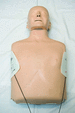
Figure 12.69
Biaxillary position of self‐adhesive electrodes.

Figure 12.76
The pillars of patient blood management.
Source : Reproduced from Shander et al. ( ) with permission of John Wiley & Sons.

Figure 12.70
Sizing an oropharyngeal airway.

Figure 12.1
The major respiratory organs in relation to surrounding structures.
Source : Marieb and Hoehn ( ).

Figure 12.5
Simple face‐mask.

Figure 12.9
Fowler position.

Figure 12.13
Heat and moisture exchanger for a ventilation circuit.
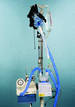
Figure 12.17
Continuous positive airway pressure (CPAP) bellows.

Figure 12.21
The relationship between the pleural membranes, chest wall and lungs.

Figure 12.25
Ambulatory chest drain bag with Heimlich valve.

Figure 12.29
Omental tag.
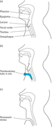
Figure 12.34
(a) Anatomy of the head and neck. (b) Tracheostomy. (c) Laryngectomy.

Figure 12.38
Cuff pressure manometer.
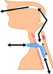
Figure 12.42
Uncuffed tracheostomy tube in situ .
Source : Reproduced with permission from the National Tracheostomy Safety Project ( www.tracheostomy.org.uk )...

Figure 12.46
(a) Jackson silver tube. (b) Negus silver tube.

Figure 12.50
Oral suction using a Yankauer suction tip.

Figure 12.54
Larnyngectomy devices. (a) Laryngecomy tube. (b) Baseplate. (c) Filter cassette. (d) Stoma covers. (e) Shower aid. (f) Tilley's forceps.

Figure 12.58
Head tilt, chin lift.

Figure 12.62
Correct hand and arm position for chest compressions.

Figure 12.66
Two people ventilating a patient using a Mapleson C system.

Figure 12.73
Vital steps involved in the transfusion of blood components.
Source : Reproduced from Bolton‐Maggs ( ) with permission of Serious Hazards of Transf...

Figure 12.77
Transfusion‐associated circulatory overload (TACO) safety checklist.
Source : Reproduced from Bolton‐Maggs ( ) with permission of Serious Hazards o...

Figure 12.71
Insertion of an oropharyngeal airway.
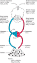
Figure 12.2
Gas movement in the body is facilitated by partial pressure differences. Top of figure illustrates pressure gradients that facilitate oxygen (O 2 ) an...

Figure 12.6
Reservoir mask (non‐rebreathe mask).

Figure 12.10
High‐flow oxygen via a nasal cannula.

Figure 12.14
Positive end‐expiratory pressure (PEEP) valves.

Figure 12.18
Bacterial–viral filter.

Figure 12.22
Large‐bore chest drain.

Figure 12.26
Triangle of safety.
Source : Laws et al. ( ). Reproduced with permission of BMJ Publishing Group, Ltd.

Figure 12.30
H‐shaped securing tape.
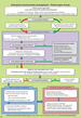
Figure 12.35
(a) Emergency tracheostomy management algorithm. (b) Emergency laryngectomy management algorithm.
Source : Reproduced with permission from the Nati...
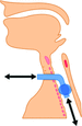
Figure 12.39
Cuffed tracheostomy tube in situ .
Source : Reproduced with permission from the National Tracheostomy Safety Project ( www.tracheostomy.org.uk ).

Figure 12.43
Portex® uncuffed fenestrated tracheostomy tubes with red inner tube to highlight that the inner tube is also fenestrated.

Figure 12.47
Rusch speaking valve.

Figure 12.51
Components of a closed‐circuit catheter. The control valve locks the vacuum on or off. The catheter is protected inside an airtight sleeve. A T‐piece ...

Figure 12.55
Red rubber tube, base plate with a heat and moisture exchanger, and a laryngectomy tube.
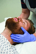
Figure 12.59
Jaw thrust.

Figure 12.63
Pocket mask with oxygen port.
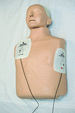
Figure 12.67
Standard position of self‐adhesive electrodes.

Figure 12.74
Cumulative data for SHOT categories 1996 to 2017, n = 19,815.
Source : Adapted from Bolton‐Maggs ( ) with permission of Serious Hazards of Transf...
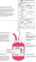
Figure 12.78
A guide to the necessary elements of blood pack labelling.

Figure 12.72
Correct placement of an oropharyngeal airway.

Figure 12.3
Oxyhaemoglobin dissociation curve. With a PaO 2 of 8 kPa and more, saturations will remain high (flat portion of curve). The middle red line is the n...

Figure 12.7
Venturi attachments.

Figure 12.11
Heat and moisture exchanger for a tracheostomy tube. (a) Swedish nose. (b) Trachphone.

Figure 12.15
Non‐invasive ventilation (NIV) patient interfaces.

Figure 12.19
Nasopharyngeal airway and safety pin.

Figure 12.23
(a) Pigtail drain with (b) magnification of the end.

Figure 12.27
Alternative positions for chest drain insertion. (a) Supine on the bed with the arm on the affected side placed behind the head away from the chest wa...
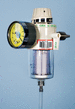
Figure 12.31
High‐volume, low‐pressure suction unit (thoracic suction adaptor).

Figure 12.36
Bed signage. (a) Tracheostomy. (b) Laryngectomy.
Source : Reproduced with permission from the National Tracheostomy Safety Project ( www.tracheosto...

Figure 12.40
Portex® Blue Line Ultra® tracheostomy tube shown with introducer.
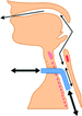
Figure 12.44
Fenestrated uncuffed tube in situ .
Source : Reproduced with permission from the National Tracheostomy Safety Project ( www.tracheostomy.org.uk )....

Figure 12.48
Passy Muir valve

Figure 12.52
Tracheostomy inner tube change.
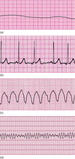
Figure 12.56
Cardiac arrest rhythms. (a) Asystole: non‐shockable. (b) Pulseless electrical activity (PEA): non‐shockable. (c) Pulseless ventricular tachycardia (VT...
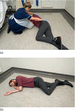
Figure 12.60
(a) Assisting the patient into the recovery position. (b) The patient in the recovery position.
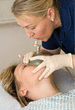
Figure 12.64
Mask with one‐way valve over patient's nose and mouth and rescuer giving breath.
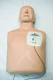
Figure 12.68
Anteroposterior position of self‐adhesive electrodes.

Figure 12.75
Bedside checklist.
Source : Reproduced from Bolton‐Maggs ( ) with permission of Serious Hazards of Transfusion.

Figure 12.79
Checking the compatibility label or tie‐on tag against the patient's wristband.