You are viewing a javascript disabled version of the site. Please enable JavaScript for this site to function properly.
Go to headerGo to navigationGo to searchGo to contentsGo to footer
Go to chapter navigation

Figure 10.1
Processing of sensory input and motor output by the spinal cord. Source : Reproduced from Tortora and Derrickson ( ) with permission of John Wiley & ...

Figure 10.5
Examples of a universal pain assessment tools. Source : Reproduced from Rhodes and Branham ( ) with permission of Pharmacy Times .

Figure 10.9
Branches of a typical spinal nerve, shown in cross‐section through the thoracic portion of the spinal cord: transverse section. Source : Reproduced f...

Figure 10.13
Patient using an Entonox demand valve.

Figure 10.17
Points used to treat migraines and headaches.

Figure 10.2
The pain pathway, showing key sites for particular analgesic interventions.

Figure 10.6
The WHO analgesic ladder.

Figure 10.10
Distribution of dermatomes.

Figure 10.14
TENS machine.

Figure 10.18
A sample acupuncture treatment record chart. Rx, prescription. Source : Adapted from BMAS ( ).

Figure 10.3
Distribution of referred pain. Source : Reproduced from Tortora and Derrickson ( ) with permission of John Wiley & Sons.
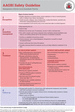
Figure 10.7
Safety guideline: management of severe local anaesthetic toxicity. Source : Reproduced from AAGBI ( ) with permission of the Association of Anaesthet...

Figure 10.11
Epidural insertion kit. Source : Reproduced with permission of Vygon.
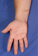
Figure 10.15
A generic acupuncture point (PC6) often used for nausea and pain.

Figure 10.4
Body diagram used for pain assessment.

Figure 10.8
Gross anatomy of the spinal cord. (a) Anterior view and transverse section through the spinal cord. (b) Transverse section of the spinal cord within a...

Figure 10.12
Entonox cylinder and hose.

Figure 10.16
A point (LI4) used for relief of pain such as headaches and toothache, for relief of sinus infections, or as a generic point in conjunction with other...